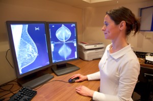
Based on a new study, an abbreviated breast MRI procedure expressed accuracy for cancer detection equivalent to that of full diagnostic MRI procedures, pointing to a possible screening method in high-risk women.
Both the abbreviated and full breast procedures detected 11 invasive breast cancers that initially went undetected by mammography" href="/tag/digital-mammography.html">digital mammography in 443 women.
“The full MRI protocol required 21 minutes to complete, as compared with about 3 minutes for the abbreviated protocol,” said Christiane K. Kuhl, MD, of the University of Aachen in Germany, at the Breast Cancer Symposium.
“The brief protocol had a negative predictive value (NPV) just less than 100%, and specificity was equivalent to that of the full protocol,” she continued. "In this preliminary proof-of-principle study, an MR table time of less than 3 minutes can help rule out breast cancer with a negative predictive values of 98.9%An MR table time of less than 3 minutes and a radiologist reading time of less than 30 seconds yielded a diagnostic accuracy that was equivalent to the regular, full diagnostic MRI protocol.”
"Abbreviated screening compares favorably with mammographic screening with regards to time needed to acquire and review images. It allowed a substantial additional yield of biologically relevant invasive cancers and ductal carcinoma in situ (DCIS) in this cohort of women at moderately or slightly increased risk of breast cancer,” she added.
Kuhl noted that breast MRI manages to identify more invasive cancers and DCIS on a regular basis than mammography does. The clinical importance of the supplementary cancers continues to be up for debate, as well as the contribution of MRI-detected cancers to overdiagnosis.
Following decades of mammographic screening, breast cancer remains a leading cause of cancer death in Europe and the U.S. in women younger than 50, Kuhl mentions.Furthermore, breast cancer remains the leading source of life-years lost in women.
Mammography identifies breast cancer on the basis of architectural distortions, spiculations, and calcifications, which echo regressive fluctuations such as hypoxia, necrosis, and fibrosis. As a result, mammography has a detection bias toward slow-growing cancers, Kuhl explained.
However, MRI detection of breast cancer is determined by angiogenic and protease activity, tissue alterations that have a direct association with carcinogenesis, cell proliferation, and metastatic growth.
"MRI detection of cancers and DCIS is biased toward biologically active, prognostically relevant disease," said Kuhl.
In order to verify such a claim, Kuhl referenced a study expressing mammography's sensitivity for DCIS decreases as grade increases. MRI's sensitivity began at 80% for low-grade DCIS (in contrast with 61% for mammography) and increased to 98% for high-grade DCIS with or without necrosis.
Recognizing cost and limited availability as restrictions on the use of breast MRI, Kuhl explained that breast MRI procedures for screening and diagnosis are quite similar. The utilization of MRI as an accurate screening system would call for reducing acquisition time and reading time, as well as dependency on radiologists with wide-ranging experience reading MR images.
Her team performed a study aiming to determine the possible exchange in MRI sensitivity with use of an abbreviated protocol appropriate for screening uses. The study included 443 women with an increased risk of breast cancer and negative digital mammograms. The patients received a total of 606 MRI studies.
Radiologists whose expertise lie in breast MRI reading carried out three readings for each MRI study: the first post contrast-subtracted images (FAST), maximum intensity projection (MIP), and results of the complete breast protocol.
The full protocol took 21 minutes to complete, including reading. FAST and MIP took 3 minutes for image attainment, 2.8 seconds for MIP reading, and 28 seconds for FAST reading
Investigators discovered 11 cancers missed by mammography (an additional yield of 18.3 per 1,000). All tumors were Tis or T1 cancers with no nodal involvement or distant metastases. MIP images were positive in nine out of 11 cases (82%), and the FAST and full-protocol studies were positive in 11 out of 12 cases (91%).
While NPV for MIP readings was 99.6%, which slightly rose to 99.8% with FAST images. FAST imaging attained a sensitivity of 94.4%, similar to the full protocol, as 33 false-positives occurred with FAST and 35 with the full protocol.
“The results could be interpreted as suggesting that DCIS detected by MRI is different from DCIS detected by mammography, “said Monica Morrow, MD, of Memorial Sloan-Kettering Cancer Center in New York City.
Kuhl stated that the lesions weren’t so different, but that MRI has a detection prejudice against clinically unimportant DCIS. Morrow questioned the legitimacy of using low-grade DCIS as a proxy for "not important."
"We have to keep in mind that every single prospective randomized trial of DCIS -- radiotherapy versus not -- showed that the risk of progression to invasive cancer was equal, regardless of the grade of DCISI'm not sure that, in and of itself, is a surrogate,” said Morrow.
