
The National Institute of Standards and Technology (NIST) has produced model modification tools for an experimental medical imaging method that provides new advantages in diagnosing and monitoring of certain cancers and potentially other medical conditions.
NIST designed, built, and tested two model phantoms for calibrating ultralow-field (ULF) magnetic resonance imaging (MRI) systems. Phantoms are frequent and widely applied tools for quality control in medical imaging. They are usually objects with simple shapes, but very well-defined responses to a particular kind of imaging scanner. As their name suggests, phantoms are stand-ins for the body, and are utilized to help optimize MRI machines to deliver the best possible medical images for a given type of tissue.
The NIST preliminary models are the first standard calibration tools for ULF-MRI, providing a quantitative way to evaluate performance, authenticate the technique, and directly compare different experimental and clinical MRI scanners.
"Tissues that may look the same in clinical MRI can look very different in ULF-MRI, which provides new contrast mechanisms. Our hope is that we can move this technique along to attract more interest from [industry] vendors,” said NIST physicist, Michael Boss.
MRI noninvasively acquires images of soft tissues predicated on measurements of how hydrogen nuclei, in the water that makes up much of the body, react to magnetic fields. ULF-MRI improves tissue contrast in particular types of MRI scans. Prostate tumors, for instance, can be quite hard to see with standard MRI, but apprear visibly clear under ULF-MRI. ULF-MRI has also been used experimentally to image the brain, and tested in at least one nonmedical application, inspection of liquids at airports.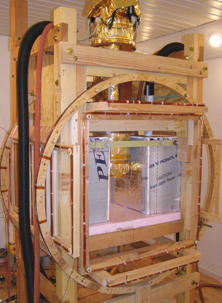
ULF-MRI also provides practical usages: The instruments are simpler in design, lighter in weight, and less costly than conventional MRI scanners. That's because ULF-MRI functions at a much lower magnetic field strength, measured in microteslas, thousands of times lower than standard MRI, which functions at up to 3 teslas and calls for huge magnets.
The low magnetic field strength means ULF-MRI requires the most sensitive magnetometers available: SQUIDs (superconducting quantum interference devices). This is fitting because it makes ULF-MRI appropriate for joining with other SQUID-based imaging techniques such as magnetoencephalography.
NIST staff previously designed phantoms for standard MRI systems and also have broad experience both making and using SQUIDs. NIST's new ULF-MRI phantoms are short plastic cylinders, shaped like hockey pucks but slightly smaller, containing six or 10 plastic jars filled with different salt solutions that become magnetized in a magnetic field. Each phantom measures a different facet of scanner performance such as spatial resolution.
NIST researchers tested the new phantoms on both a standard MRI system at the University of Colorado Health Sciences Center (Denver, Colo.) and an experimental ULF-MRI scanner at the University of California (UC) at Berkeley, where the method was first presented about a decade ago.
Tests results indicate the prototype phantoms are well-suited to ULF-MRI applications and allow direct comparison of ULF and clinical MRI system performance.
NIST researchers now plan to integrate design improvements predicated on lessons learned from the prototypes, with the goal of enhancing phantom stability and providing traceability to standard measurement units. NIST and UC Berkeley researchers also plan to collaborate to further develop ULF-MRI technology for detection of prostate and breast cancers.